Retinal
Photoisomerization of Retinal Chromophores

Retinal is the photoactive component in various biologically relevant receptor proteins. It triggers perception, ion pumping or energy production in rhodopsins, a family of seven-helical transmembrane proteins. The initial reaction features a light induced change of the chromophore configuration: retinal isomerizes from 11-cis to its all-trans form in mammalian rhodopsin, whereas archaeal rhodopsins invole all-trans to 13-cis photoisomerization. This initial step takes place on the femtosecond timescale and drives further, thermally activated rearrangements in the protein to fulfill its function. We investigate the initial and ultrafast photoisomerization of retinal chromophores in their protein environments using combined quantum mechanical/molecular mechanical (QM/MM) approaches. With atomistic molecular dynamics simulations using the COBRAMM software,1 we follow the motions of the molecules on the excited and ground state potentials to obtain information on reaction channels, product distributions (i.e. quantum yields) and reaction times
Bovine and human rhodopsin

Vision in vertebrates is steered by 11-cis to all-trans photo-isomerization of retinal with ca. 65% quantum yield. The first spectroscopically detectable photoproduct is batho-rhodopsin, where the chromophore entails a twisted all-trans configuration. The twists store energy, which is released in later thermal steps. A series of biochemical reactions finally lead to the excitation of the optical nerve. With CASSCF excited state trajectories including surface hopping,2 we were able to follow the molecular motions and spectral traces of retinal during this photoprocess. The reaction involves a unidirectional bicycle-pedal-like motion of the C9=C10 and C11=C12 double bonds. The hydrogen-out-of-plane modes at the isomerizing double bond play a central role in the generation of the photoproduct. Bi-directional deactivation was found in the analogue iso-rhodopsin protein, that has a 9-cis chromophore.3
Channelrhodopsin
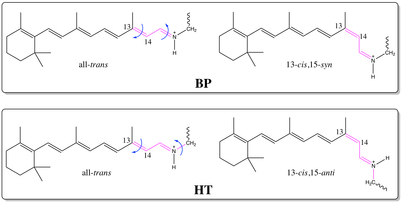
Unlike in mammalian rhodopsins, where a complex biochemical cascade provides signaling, the channelrhodopsins (ChRs) found in certain types of green algae directly respond to a stimulation by light. Upon excitation and all-trans to 13-cis isomerization of retinal, a channel opens in the membrane that quickly depolarizes the cell. This property and the fact, that ChRs can be easily expressed into living tissue opened a new field in neuroscience. Optogenetics uses light-activated proteins to influence cell functionality. Optogentical tools can e.g. be used to study nerve-regulation functions non-invasively by light irradiation. To modify the properties of ChRs for a wider application, a detailed understanding of the initial photoisomerization is necessary. We use the OMx/MRCI method and the Amber force field to simulate the photoreaction of retinal in the chimeric C1C2 protein.4 The identified reaction channels feature bicycle-pedal (BP) and hula-twist (HT) motions. These motions lead to different orientations of the NH+ proton of the retinal chromophore, that is transferred to one of the two negatively charged counterions ASP and GLU in a later thermal step. The spectroscopic and dynamics properties of retinal are largely modified by these counterions.5
References
- Weingart, O. et al. COBRAMM 2.0 - A software interface for tailoring molecular electronic structure calculations and running nano-scale (QM/MM) simulations. J. Mol. Model. DOI: 10.1007/s00894-018-3769-6 (2018).
- Polli, D. et al. Conical intersection dynamics of the primary photoisomerization event in vision. Nature 467, 440-443 (2010).
- Polli, D. et al. Wavepacket Splitting and Two-Pathway Deactivation in the Photoexcited Visual Pigment Isorhodopsin. Angew. Chemie Int. Ed. 53, 2504-2507 (2014).
- Dokukina, I., Nenov, A., Marian, C. M., Garavelli, M. & Weingart, O. QM/MM photodynamics of Retinal in the Channelrhodopsin Chimera C1C2 with OM3/MRCI. ChemPhotoChem submitted, (2018).
- Dokukina, I. & Weingart, O. Spectral properties and isomerisation path of retinal in C1C2 channelrhodopsin. Phys. Chem. Chem. Phys. 17, 25142-25150 (2015).